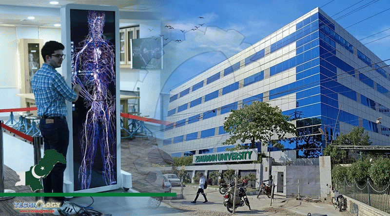
The Ziauddin University in Karachi has introduced a 3D virtual dissection table, first of its kind in Pakistan, at its new digital anatomy lab.

The Ziauddin University in Karachi has introduced a 3D virtual dissection table at its new digital anatomy lab in Clifton. It is reportedly the first of its kind in the country.
“It’s the very first time in the history of Pakistan that Ziauddin University [is] introducing the 3D virtual dissection table which is being taken to all the campuses of Ziauddin University and hospitals to help students in getting technology-based learning,” Ziauddin University Chancellor Dr Asim Hussain said at the inauguration Thursday.
The new dissection table, called Anatomage, will be used as a learning tool for university students of all years. It can be connected to different physical locations across all three campuses.
“It will be helpful for all medical and dental students, labs, radiologists, nurses, surgeons, doctors and house officers,” said Dr Bushra Wasim Khan, a professor and head of the Anatomy Department at Ziauddin University.
Anatomage uses the latest 3D technology to allow users to visualise every structure of the body in great anatomic detail.
“Your finger becomes the scalpel,” Dr Abbas Zafar, a professor and dean of Ziauddin Faculty of Health Sciences, told SAMAA Digital.
“You can visualise and rotate every organ at different angles and look at any cross-section.”
With the touch of a finger a tissue layer can be peeled to reveal underlying structures.
In our times, we had to work on cadavers — the scientific term for corpses used for medical dissection — in hot, crowded rooms, Dr Zafar added.
Most of these cadavers were the unclaimed bodies brought from hospital morgues. The practice largely died down when the Supreme Court banned the use of real human bodies in medical schools.
The bodies were preserved using formalin which is a pungent chemical and can be toxic and allergenic. The experience was often emotionally disturbing for students.
“Now you can carry out dissection in a congenial atmosphere with no unpleasant smells,” Dr Zafar said.
Medical dissection was replaced by physical 3D models of the body. These, however, were plastic and lacked realistic detail. Learning wasn’t immersive the way it can be with the 3D visual dissection table, according to the professor.
It is the only fully segmented real human 3D anatomy system, according to Ziauddin University. This means any section cut through virtually will look exactly like it would in the body.
The technology was invented by Stanford professors in the 80’s. They reconstructed the human body using cross-sections of real bodies, mapping all blood vessels, nerves and their connections.
Anatomage is compatible with X-rays, CT scans and MRI, allowing detailed radiology reviews for students. They will be able to view how different disease processes affect the organs and how they would show up in imaging.
It is already widely used in the US and Europe. Research has proven that virtual dissectors can help improve student scores.
Originally published at SAMAA