The findings may have neutrons applications in drug development, and they also address long-standing fundamental mysteries about why cell membranes move as they do.

We now have a clearer picture of the lightning-fast molecular dance occurring within the membrane that encloses each cell in our body, revealed in part by neutron beams at the National Institute of Standards and Technology (NIST). The findings may have applications in drug development, and they also address long-standing fundamental mysteries about why cell membranes move as they do.
The research, published today in Physical Review Letters, provides new insight into Neutrons how the movements of the individual lipid molecules that form the membrane affect its overall properties—particularly its viscosity, or resistance to flow. Understanding these properties is important because the membrane—the boundary between the cell and its surroundings—holds the key to accessing its interior.
“We discovered the time scale at which the lipid molecules are moving, and we connected it with the membrane’s viscosity,” said Michihiro Nagao, a scientist at NIST and the University of Maryland who performed the work with his colleagues at the NIST Center for Neutron Research (NCNR). “We have evidence of where the viscosity comes from, and we also show that our tools can study it. We didn’t have an effective technique to explore it before, so it’s an important advance.”
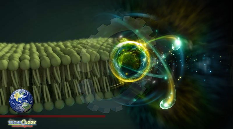
While the membrane is a nominally solid barrier between the cell and its surroundings, the fatty lipid molecules that form it alternately embrace, glide and repartner themselves continually, making the membrane act more like a sticky, viscous fluid such as honey or oil. Suspended in the membrane are membrane proteins and transport channels that function as gateways to the cell’s interior. Until recently, though, it was difficult to study the lipid molecules effectively because they move so quickly that their dance was hard to follow.
“Trying to understand how the protein channels work without considering the membrane is like trying to understand a fish without considering water,” said NIST’s Elizabeth Kelley. “We wanted a better perception of how the lipids move.”
Visualizing those movements is now possible by probing them with neutrons at the NCNR and X-rays from Japan’s SPring-8 synchrotron. Scientists from the two facilities collaborated to get the results. They first created a model membrane of lipid molecules, each of which has a bulbous head that forms the membrane’s outer surfaces and two tails that form its interior. The lipids were essentially identical to those in natural cell membranes, with the exception that all the hydrogen atoms were replaced by deuterium, which shows up more clearly in neutron scans.
A membrane, which is only two molecules thick, is essentially a two-dimensional sheet of oil, making it difficult to research its viscosity as it moves. While it is easier to research 3D oils, past attempts to estimate the viscosity of 2D lipid membranes from the viscosity of the corresponding 3D oil haven’t worked well. The new findings indicate that packing the lipids into a membrane slows their motions and increases the interactions between molecules, leading to a higher viscosity than a 3D fluid would have by Neutrons.
The neutron beams helped the team explore two types of molecular motion that relate to membrane viscosity. One type concerned the movement of the tails in the model membrane. The tails, which are tightly packed in an even thinner layer between the lipids’ heads, move very fast, quivering once every 10 picoseconds, or trillionths of a second. While these motions are incredibly quick, they are actually an order of magnitude slower than scientists have predicted from the motions in a 3D liquid oil, suggesting that the 2D membrane structure and interactions between the lipids are key to determining its viscosity.
The other type concerned the movement of the full lipid molecules as they danced around one another within the membrane. The molecules, it turns out, move about 10 times slower than their tails do. The friction the molecules experience, combined with the friction among their tails, produces a viscosity measure that falls in the middle of the range of viscosity estimates past research efforts have indicated—suggesting the measurements account for all the factors that contribute to the viscosity.
“It’s a combination of sources of friction on the molecules that creates the membrane’s viscosity,” Nagao said. “You need to consider the tails contacting one another, the full molecules rubbing against one another and some other factors such as the heads interacting with the water around them. But if you put all the sources together, you get a viscosity measurement that accords well with previous estimates.”
Much of the experimental data was obtained using the neutrons spin echo spectrometer, one of five CHRNS instruments that are partially funded by the National Science Foundation to help explore materials. The molecular-scale motions it revealed are relatively easy to study using computer simulation techniques, meaning that the fundamental knowledge the experiment provided might help improve these computations and thus aid drug discovery.
“Measuring the viscosity helps us understand how quickly things move around in the membrane and how long it takes to open the cell,” Kelley said. “These sorts of insights may help us design drugs that take advantage of them.”
Source Phys.Org